MORPHOLOGY AND SURFACE CHARACTERIZATION
| Technique | Type of apparatus |
Partner name, Contact person Expertise of the personel |
Types of possible experiments |
| Scanning Force Microscopy |
Multimode 8, NanoScope-D3100, NanoScope V-D3100, Dimension ICON / MFP-3D |
IPF
Dipl.-Phys. Andreas Janke Tel.:+49(0)3514658-496
surface characterization by AFM (since 1992) and SEM
|
- Surface morphology / roughness - Phase imaging / material contrast, electrical charge distribution, magnetic contrast - Mechanical Properties: E-modulus, adhesion, energy dissipation |
| SFM force measurements | NanoScope IIIa-D3100, NanoScope V-D3100, Dimension ICON, Multimode 8 |
IPF
Dr. rer. nat. Astrid Drechsler Tel.: +49(0)3514658-540
Polymer surfaces: morphology, roughness, interaction forces, contact angle, wetting and absorption, adsorption
|
- Investigation of surface morphology, roughness, material contrast - Force measurements in air and fluids at variable temperature - Colloidal probe technique |
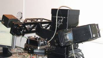
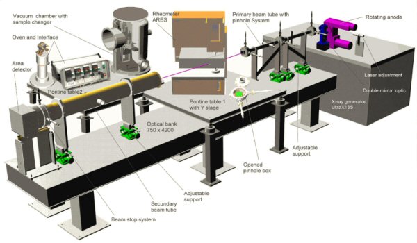
| RheoSAXS (WAXS) Combination of SAXS / WAXS with rheometer or mechanical tensile testing Combination of SAXS / WAXS geometry with heating chamber / mechanical tensile testing |
3-pinhole collimated X-ray camera equipped with rotating anode generator, confocal optic and area detector MarCCD |
IPF
Prof. Manfred Stamm, Tel.:+49(0)3514658-224 Dr. Dieter Jehnichen Tel.:+49(0)3514658-493 Investigations in polymers and polymer nanomaterials
|
- Structureand structure changes in polymers and polymer nanomaterials - Order-disorder transitions of nanostructured polymers |
| 4-circle wide-angle diffractometer P4 | WAXS, Karlsruhe, Germany [former: Siemens] |
IPF
Dr. Dieter Jehnichen Tel.:+49(0)3514658-493 -Polymer physics, physical chemistry of polymers -Structure-property relationships |
WAXS (and IMAXS) in transmission technique -2D-scattering pattern of bulky material, powder, fibres or otherwise oriented (textured) samples, determination of crystallinity, crystallite size, orientation parameters -Phase analysis (determination of modificatons) |
| SAXS-Kratky |
KRATKY compact small-angle system with temperature control [Hecus, Graz,Austria] |
IPF Dr. Dieter Jehnichen Tel.:+49(0)3514658-493 |
- SAXS (and IMAXS) in transmission technique Morphology of standard polymers -nanocomposites as to the intercalation and exfoliation processes |
| XR/GID/WAXS (Θ/Θ-diffractometer) |
2-circle X-ray diffractometer XRD 3003 T/T [GE Inspection Technologies, Ahrensburg, Germany |
IPF Dr. Dieter Jehnichen Tel.:+49(0)3514658-493 |
- WAXS (and IMAXS) - Crystallinity, crystallite size, coefficients of linear expansion -phase analysis - Simple depth profile - Temperature-dependent experiments for discontinuous heating/cooling cycles |
|
D8 DISCOVER diffractometer system for WAXS/IMAXS and XR/GID |
Vertical 2-circle goniometer with Bragg-Brentano or parallel beam geometry (pinhole or slit collimated) and 2D or 1D detector equipped with Euler cradle and temperature unit [Bruker AXS Karlsruhe, Germany] |
IPF Dr. Dieter Jehnichen Tel.: +49(0)3514658-493 Polymer physics, physical chemistry of polymers -Structure-property relationships. Investigations in polymers, nanomaterials, composites and blends |
- Structure and structure changes of bulk material, fibers and thin films - Crystallinity, crystallite size, coefficients of linear expansion, phase analysis, orientation parameters - Depth profile, roughness - Temperature-dependent experiments for discontinuous heating/cooling cycles |
| Ellipsometer |
Spectroscopic ellipsometer M-2000VI |
IPF Dr. Klaus-Jochen Eichhorn Tel.:+49(0)3514658-256 Ellipsometry measures in polymers. |
- Thickness and optical constants of thin layers on solid substrates - In situ experiments on the solid/liquid interface |
| Contact angle, surface tension |
OCA 40 micro, OCA 35XL (Dataphysics) Fibro Dat 1100, ADSA-P, OBS2, OBS3 (in-house developments) Tensiometer K12, K14 (Kruess GmbH), DCAT21 (Dataphysics) |
IPF Dr. Karina Grundke Tel.:+49(0)3514658-475 Characterization of polymer interfaces, wetting and interfacial tension of polymers (including polymer melts), drop profile analysis, interfacial phenomena in multi-component systems (coatings, reinforced polymer composites, biomaterials, microelectronic applications, interrelations between adhesion, wetting and adsorption) |
- Contact angle measurements (sessile drops, captive bubbles, indirect measurement by the Wilhelmy technique, advancing and receding contact angles, capillary penetration of liquids into porous solid systems, e.g. powder packings, fiber bundles, membranes, filters); liquid surface tension including polymer melts at elevated temperatures and simultaneous measurement of surface tension and density of polymer melts; Approaches to estimate the surface energetics of polymers (surface free energy, thermodynamic work of adhesion) from wetting and surface tension measurements |
| Optical roughness analysis (MicroGlider) | MicroGlider (chromatic white light sensor) and Thin Film Sensor FTR (VIS-interferometer), Fries Research and Technology GmbH | IPF Dr. rer. nat. Astrid Drechsler Tel.: +49(0)3514658-540 Polymer surfaces: morphology, roughness, interaction forces, contact angle, wetting and absorption, adsorption |
- Quantitative determination of surface morphology and topometry, roughness and waviness on microscopic scale - Thickness determination of e.g. polymer layers using optical non-contact methods |
| Confocal microscopy | µsurf explorer (scan-disk confocal high resolution microscope), NanoFocus AG | IPF Dr. rer. nat. Astrid Drechsler Tel.: +49(0)3514658-540 |
- Quantitative topographic surface characterisation; investigation of micro-defects |
| Non-contact surface profilometry | TRACEiT, INNOWEP GmbH Measuring & Testing | IPF Dr. rer. nat. Astrid Drechsler Tel.: +49(0)3514658-540 |
- Spectrophotogrammetry: analysis of topography and morphology of rougher surfaces. - Translucency of materials |
| Micro-mechanical and tribological analysis | Universal Surface Tester (UST), INNOWEP GmbH Measuring & Testing | IPF Dr. rer. nat. Astrid Drechsler Tel.: +49(0)3514658-540 |
- Analysis of micro-mechanical, micro-tribological and functional properties of materials in situ in the submicrometer range using abrasion, wear, scratch resistance, micro friction, structure and haptic parameters - Evaluation of surface mechanical properties of plastics, lacquers, coatings, fabrics, polymers, metals, ceramics, rubber and biological materials. |
| Dynamic contact angle and absorption measurement |
DAT1100 dynamic contact angle tester with DAT 1129 automatic tilt table, FIBRO system AB, Sweden |
IPF Dr. rer. nat. Astrid Drechsler Tel.: Tel.: +49(0)3514658-540 |
- Dynamic contact angle, wetting and absorption measurements on porous and nonporous materials. - Characterisation of textiles, nonwovens, papers, powders, membranes etc. |
| Electrokinetic analyzer EKA |
Producer: A. Paar | IPF Dr. Cornelia Bellmann Tel.:+49(0)3514658-327 colloid chemistry, solid surface analysis |
- Streaming potential measurements vs. pH, time, electrolyte concentration, surfactant or polyelectrolyte concentration |
| Acoustic and electroacoustic spectrometer DT-1200 | Dispersion Technology, Inc. | IPF Dr. Cornelia Bellmann Tel.:+49(0)3514658-327 colloid chemistry |
- Electroacousticstudies for calculation electrokinetic data - Study of particle size distribution by acoustic attenuation measurements |
| Microscopy | Optical and stereo microscopes | SICOMP Lars Liljenfeldt, Marketing manager Tel.: +46 (0) 911 744 40 |
- Measuring of void content - Measuring of crack propagation - Surface characterisation |
| NDT-testing | C-scan, Sonatest, RapidScan2 | SICOMP Lars Liljenfeldt, Marketing manager Tel.: +46 (0) 911 744 40 |
- Quality control with respect to voids, delamination and other defects |
| 3D-scanning | ARAMIS, by GOM, 3D-deformation measurements ATOS, by GOM, 3D-scanner for geometry measurements |
SICOMP Lars Liljenfeldt, Marketing manager Tel.: +46 (0) 911 744 40 |
- Deformation measurements - 3D-geometry measurements of components and tooling |
| Scanning Force Microscopy | Solver PRO, NT-MDT | TUL Dr. Izabela Bobowska Tel +48 426313205 |
- Surface morphology / roughness - Phase imaging / material contrast, electrical charge distribution, magnetic contrast |
| Raman Spectrometer with confocal microscope | T 64000 Jobin-Yvon |
TUL Dr. Marcin Kozanecki Tel: +48 426313205 Micro-Raman analysis of heterogeneous systems (organic, inorganic, hybrid) -Low Frequency Raman Scattering |
- Micro-Raman study with back scattering configuration using confocalmicroscope: - depth profiling - mapping - Polarized Raman spectra |
| FEG-SEM Microscopy |
Field emission scanning electron microscope (FESEM, Leo Supra 35) equipped with Energy dispersive X-ray spectroscopy (EDX INCA, Oxford Instruments, UK). |
INSTM research Unit University of Rome “Tor Vergata” Prof. Francesca Nanni Tel: +39 0672594496 |
electron images of conductive and poorly conductive surfaces, chemical microanalysis (qualitative and quantitative) of samples. Sputter coater for the preparation ad observation of non-conductive samples |
| Optical Microscopy |
Optical microscope (NIKON SMZ-U) |
INSTM research Unit University of Rome “Tor Vergata” Prof. Francesca Nanni Tel: +39 0672594496 |
Observation of samples at different magnification (up to 75x), digital camera to capture images for subsequent analysis |
| Optical Microscopy | Metallographic Microscope (NIKON Epiphot Japan 46773) and Apparatus for metallographic sample preparation (Sectioning and Cutting machine ISOMET 4000 Buhler, Polishing machine, etc. |
INSTM research Unit University of Rome “Tor Vergata” Prof. Francesca Nannii Tel: +39 0672594496 |
Microstructure analysis of metallographic prepared specimens at different magnifications (up to 1000x) |
| X-Ray Diffractometry | X-ray diffractrometer (XRD, Philips X Pert) equipped with high-temperature device (HT-XRD, Tmax 1100 °C) (Anton Paar HTK 1200) and accessories for powders, bulk and thin film samples |
INSTM research Unit University of Rome “Tor Vergata” Prof. Francesca Nanni Tel: +39 0672594496 |
Analysis of phases of bulk materials, powders, films and electrospun mats. Database of XRD charts for phase analysis. Determination of residual stress of coatings. Phase analysis under heating up to 1100°C. |
| Polarized Optical Light Microscopy | Polarized Optical Light Microscopy, Hund mod. H600, |
INSTM Prof. J.M. Kenny |
Study of crystals growth by means of a coupled Mettler hot stage |
| Contact Angle |
FTA2000 First Ten Angstroms |
INSTM Prof. J.M. Kenny |
Measurement of contact angle – surface tension, surface energy, work of adhesion, static, advancing/receding contact angles, pendant drop and oscillating drop surface tension. |
| Atomic Force Microscope |
AFM System, NanoSurf Easy Scan, Swiss STM, Easy Scan, Nanosurf, Swiss |
INSTM Prof. J.M. Kenny |
Carbon nanotubes alone or in nanocomposite, organic and inorganic polymers (metalized), stem cells on polymers, antique ceramics, epoxy resins, proteins Topography of surfaces |
| Shearing Hot Stage for Microscope | CSS 450Linkam | UCBL Dr. René Fulchiron -Flow induced crystallization of polymers -Morphology development after shear in polymer blends |
- Application: Polymer crystallization, polymer blends, droplet deformation. |
| Scanning Electron Microscopy | Environmental scanning electron micrsocope |
CSIC José David Gómez, Technical Engineer Pattern surfacecharacterization |
- Surface characterization - Measurements or surface patterns, images of surface details. |
| FTIR-ATR |
Spectrum One Perkin Elmer/ Pike Technologies |
CSIC Dr.Helmut Reinecke Surface selectivities in polymericfilms |
-Structural information on liquids and solid surfaces, - Qualitative depth profiles on solid surfaces |
| Plasmon Resonance Spectroscopy | α-SPR prototype, Sensia SL |
CSIC Dr.Alberto Gallardo |
- Biomaterial characterization, since it is able to monitor in real time and in situ any dynamic process |
| Scanning Force Microscopy | NANOSCOPE Multimode Digital Instruments (VEECO) |
TECNALIA José Luis Viviente, researcher |
-Biomaterials and biomedical fields. -Bionanotechnology and nanomaterials -Microandnanometricdimensionalmeasurements. -Electric potential and force measurements |
| Auger Electron Spectroscopy (AES) / X-Ray Photoelectron Spectroscopy (XPS) |
MICROLABMKII V.G.Scientific |
TECNALIA José Luis Viviente, researcher -Biological, biomaterials and biomedical fields. -Nanotechnology, bionanotechnology and anomaterials. |
Characterization of solid samples surfaces extracted from different materials electrically conductive: metals, ceramics, composites, fibres, materials with different surface treatments, thin films of greasy or other compounds -determination in a superficial level (first atomic layers ~3 nm) |
| Electron microscopy | High resolution SEM microscope (Quanta 200 FEG), Standard TEM microscope (Tecnai G2 Spirit 120kV) Equipment for preparation of polymer samples (ultramicrotomy, staining…) |
IMC Prague Dr. Miroslav Slouf Tel.: +420 296809291 Polymer morphology |
- SEM and TEM microscopy of bulk polymer samples including sample preparation (fracturing, ultraghin sectioning, staining, etching…) - SEM and TEM microscopy of nanoparticles (dried – standard TEM and STEM, in water – wetSTEM, frozen – cryoTEM) - EDX analysis (in both SEM and TEM) - EM analysis of polymer hydrogels (samples with high water content – cryoSEM, ESEM) |
| Stylus Profiler |
DektakXT-E, Bruker |
TUL Dr Ireneusz Glowacki Tel.: +48 426313205 |
- Surface morphology / roughness - Contact measurement |
